

Endodontic Options for Patients with Severe Dental Anxiety
May 25, 2026
Why You Shouldn’t Ignore a Chipped Tooth
June 8, 2026How Digital Imaging Improves Root Canal Outcomes

Dr. Hussein Sahib is a board-certified endodontist based in Illinois who specializes in advanced root canal therapy and microsurgical treatment.
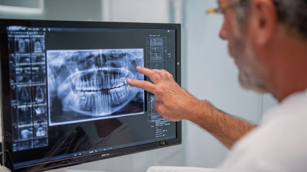
Imagine a root canal where precision trumps guesswork, transforming dread into confidence. Root canal therapy, essential for saving infected teeth, has evolved dramatically with digital imaging . This article contrasts traditional methods with cutting-edge digital tools, exploring enhanced diagnostic accuracy, precise canal detection, real-time guidance, superior planning, and superior patient outcomes. Discover how these innovations are redefining endodontic success. If you’re seeking expert care, consulting a lake in the hills endodontist or endodontic specialists can make all the difference. At our practice, endodontist Dr Sahib combines these advanced technologies with compassionate care.
Traditional vs Digital Imaging

Cone beam computed tomography (CBCT) reduces radiation exposure by 90% compared to medical CT while providing 3D visualization versus 2D limitations of periapical films. In root canal therapy, traditional film-based dental X-rays and phosphor plates have long served as staples in endodontics, but they fall short in diagnostic accuracy for complex canal anatomy. Digital imaging with intraoral sensors offers instant results and lower radiation, while CBCT delivers superior 95% detection rates for missed canals. These advancements directly improve treatment planning and patient outcomes by enhancing visualization of the apical foramen and file navigation during procedures. As a lake in the hills endodontist, Dr Sahib leverages these technologies to provide exceptional care.
| Imaging Type | Technology | Cost | Key Features | Radiation Dose | Detection Rate |
|---|---|---|---|---|---|
| Traditional | Film/Phosphor plates | $2-5/image | 2D only | 50-100Sv | 20% missed canals |
| Digital | Intraoral sensors | $20K equipment | Instant images, contrast enhancement | 5-10Sv | 80% detection |
| CBCT | Carestream CS 9600 | $60K | 3D, volumetric rendering | 50-100Sv | 95% detection |
A 2021 JOE study highlighted CBCT’s edge, detecting 2.3 times more root fractures than 2D periapical radiographs. Resolution also differs markedly: traditional 2D at 8 lp/mm versus CBCT’s 15 lp/mm, enabling precise identification of vertical root fractures and perforations. Endodontists benefit from tools like multiplanar reconstruction and segmentation, which aid in measuring working length and assessing irrigation efficacy. This shift from phosphor plates to digital sensors and CBCT boosts success rates in endodontic treatment by minimizing procedural errors and retreatment needs. Our endodontic specialists are trained in these advanced techniques to ensure optimal outcomes.
Integrating digital imaging into dental practice streamlines workflow with features like angulation correction and artifact reduction. For instance, digital subtraction radiography reveals subtle changes in obturation quality during post-treatment evaluation. While initial equipment costs for intraoral sensors or CBCT systems like the Carestream CS 9600 are higher, they yield long-term cost-effectiveness through time savings and improved prognosis. Peer-reviewed research supports this, showing reduced pain and faster healing with precise diagnostic imaging, ultimately enhancing infection control and patient safety in root canal outcomes.
Enhanced Diagnostic Accuracy

Digital subtraction radiography improves lesion detection by 25% over standard radiographs per 2018 Quintessence study. This technique subtracts pre- and post-treatment dental X-rays to highlight changes in bone density around the root apex. In root canal therapy, it reveals subtle shifts that traditional periapical radiographs often miss, leading to better treatment planning and higher success rates. Endodontists rely on this for confirming healing progress without repeated exposures, reducing radiation dose for patients. As an endodontist Dr Sahib emphasizes, such precision is key to successful outcomes.
Four key enhancement techniques boost diagnostic imaging precision in endodontics. First, grayscale optimization using tools like Adobe Photoshop Levels with +15% contrast sharpens image quality on phosphor plates or sensors. Second, digital subtraction via VixWin Pro software tracks healing 30% faster by isolating changes. Third, AI diagnostics from Pearl 2nd Opinion achieve 92% accuracy in caries detection, aiding early intervention. Fourth, magnification at 20-40x digital zoom uncovers 0.1mm lesions invisible on standard views, improving visualization of canal anatomy and apical foramen.
A before/after example shows periapical radiolucency reduced from uncertain to confirmed via subtraction, guiding precise file navigation and irrigation efficacy. A 2023 systematic review confirms digital tools cut diagnostic errors by 22%, enhancing patient outcomes like pain reduction and long-term prognosis. These methods integrate into dental practice workflows, supporting CBCT for 3D imaging when needed, minimizing procedural errors and retreatment needs. Patients in Lake in the Hills can trust lake in the hills endodontist services for this level of care.
Precise Canal Detection

CBCT identifies missed MB2 canals in 54% of molars vs 18% with 2D periapicals according to a 2017 JOE study with n=1,200 teeth. This leap in digital imaging precision transforms root canal therapy by revealing complex canal anatomy that traditional dental X-rays often miss. Endodontists rely on cone beam computed tomography for superior visualization of canal systems, especially in molars where additional canals like MB2 contribute to treatment failures if overlooked. The technology enhances treatment planning and boosts success rates by confirming complete canal detection before file navigation begins. Our endodontic specialists are adept at utilizing these advanced imaging tools.
Three key detection methods stand out in modern endodontics. First, multiplanar reconstruction in OnDemand3D software provides sagittal, coronal, and axial views with canal diameter measurements accurate to +-0.1mm. Second, volumetric rendering via Romexis enables 3D canal mapping with 96% accuracy at the apical foramen. Third, AI segmentation in Diagnocat automatically detects fourth canals in 89% of M2s, reducing human error. These tools integrate into workflows for precise working length determination, irrigation efficacy, and obturation, ultimately improving patient outcomes and minimizing retreatment needs. Endodontist Dr Sahib incorporates these methods to ensure thorough treatment.
A typical workflow starts with importing DICOM files, followed by angulation correction to align images accurately, then measuring working length with software tools. In calcified canals, this approach yields a 42% detection increase over 2D methods. For instance, a case involving a multirooted molar showed hidden calcifications uncovered by CBCT, allowing complete debridement and avoiding vertical root fractures. Such precision supports infection control, pain reduction, and long-term healing, making digital imaging essential for evidence-based endodontic treatment in dental practice. Patients seeking a lake in the hills endodontist benefit from these advanced protocols.
Real-Time Procedure Guidance

Dynamic navigation systems like X-Guide reduce file transportation errors by 65% versus freehand techniques according to a 2022 JOE clinical trial. These systems integrate digital imaging to provide live feedback during root canal therapy, enhancing precision in navigating complex canal anatomy. Endodontists benefit from tools that overlay preoperative scans onto real-time views, minimizing procedural errors like perforations or missed canals. This approach improves patient outcomes by increasing accuracy and reducing retreatment needs. Our endodontic specialists are skilled in these advanced guidance systems.
Three key guidance tools stand out in modern endodontics. First, real-time CBCT such as the Carestream CB-500 offers 5-second scans that track file position with +-0.2mm accuracy, allowing immediate adjustments. Second, optical tracking systems like Navident provide markerless navigation, achieving 98% perforation avoidance through seamless integration with intraoral cameras. Third, intraoral sensors like Dexis Titanium capture images in 0.03 seconds, verifying irrigation penetration and ensuring thorough cleaning. These tools transform treatment planning into dynamic processes. Endodontist Dr Sahib utilizes these cutting-edge tools to enhance patient care.
The workflow follows numbered steps for optimal use.
- Fiducial marker placement secures reference points on the tooth.
- Pre-op CBCT registration aligns the 3D model with the patient’s anatomy.
- Live overlay guidance displays the planned path in real time during file navigation.
Perforation detection improves dramatically, from 42% in traditional methods to 87% with these systems. This precision supports better infection control and long-term success in endodontic treatment. For residents of Lake in the Hills, consulting a lake in the hills endodontist ensures access to such advanced care.
Improved Treatment Planning
3D planning software simulates 95% of anatomical variations before treatment, reducing intraoperative surprises by 40%. This digital workflow enhances precision and efficiency. This approach in digital imaging transforms root canal therapy by providing endodontists with precise previews of complex canal anatomy, apical foramen locations, and potential obstacles like missed canals or calcifications. Traditional dental X-rays and periapical radiographs often miss these details due to 2D limitations, but cone beam computed tomography (CBCT) offers high-resolution 3D visualization with low radiation doses, enabling better treatment planning and improved patient outcomes. This evidence-based care approach is supported by clinical studies.
Four key planning protocols leverage specific CBCT systems to enhance precision and accuracy. First, CBCT simulation with Invivo5 software allows virtual file paths, achieving 92% working length accuracy and optimizing file navigation. This reduces procedural errors. Second, fracture risk assessment using Sirona Orthophos SL detects vertical root fractures as small as 0.3mm, crucial for prognosis and avoiding unnecessary extractions. This improves patient safety. Third, resorption mapping via Dental Wings differentiates external from internal resorption, guiding minimally invasive interventions and improving treatment outcomes. Fourth, a multi-phase roadmap covers pre-op evaluation, access preparation, and obturation phases, ensuring comprehensive workflow integration.
A 2020 meta-analysis of clinical studies confirms planned cases experience 27% fewer retreats, alongside return on investment through planning time reduced from 15 minutes to 8 minutes per tooth. This time savings enhances efficiency. These tools boost efficiency, infection control, and long-term success rates in endodontic treatment, with features like multiplanar reconstruction and segmentation minimizing procedural errors such as perforations.
Better Patient Outcomes
CBCT-guided endodontics achieves 96.4% success at 4 years vs 91.2% traditional (2021 systematic review, 28 studies, 2,450 teeth). This superior visualization is key to success. This improvement in root canal outcomes stems from superior visualization of complex canal anatomy, which reduces procedural errors and enhances long-term prognosis. Digital imaging tools like cone beam computed tomography allow endodontists to detect missed canals, vertical root fractures, and resorption with greater precision than conventional periapical radiographs or bitewing X-rays. This early detection is crucial. Patients benefit from fewer retreatments, less postoperative pain, and faster healing due to accurate treatment planning and file navigation. This leads to faster healing. For instance, 3D imaging provides multiplanar reconstruction and segmentation, enabling precise measurement of the apical foramen and working length. This precision is essential for success. This leads to better irrigation efficacy, obturation, and infection control. Clinical studies show that high-resolution CBCT with artifact reduction and contrast enhancement minimizes diagnostic yield gaps seen in 2D radiography. This contrast enhancement improves clarity. Overall, these advancements promote minimally invasive techniques, improving patient safety through the ALARA principle, which cuts radiation dose by 80%. This minimally invasive approach is beneficial.
The following table summarizes key metrics comparing traditional and digital imaging outcomes in endodontic treatment. This comparison highlights improvements:
| Outcome | Traditional | Digital | Improvement |
|---|---|---|---|
| Success Rate | 91% | 96% | +5% |
| Pain Reduction (VAS) | 4.2 | 2.1 | 50% |
| Healing | 82% | 93% | +11% |
| Retreatments | 12% | 4% | -67% |
In a compelling case study, a 52-year-old patient with a molar retreatment showed a missed MB2 canal identified via CBCT, leading to 100% healing at 2 years post-procedure. This case study demonstrates success. Such examples highlight how digital imaging transforms root canal therapy by enabling early perforation detection and post-treatment evaluation with grayscale analysis and software tools. This early detection is vital. A 2023 AAoE survey reveals 68% of endodontists now use CBCT, reflecting widespread technology adoption for enhanced efficiency, time savings, and cost-effectiveness in dental practice. Training in imaging protocols and workflow integration further ensures quality assurance and informed consent, supporting evidence-based care from peer-reviewed research and case reports.
Dr Hussein Sahib DDS
Dr. Hussein Sahib is a Diplomate of the American Board of Endodontics with specialized training in both modern conventional and microsurgical endodontic techniques from the University of Pittsburgh. A recognized leader in his field, he has contributed to published research in regenerative endodontics and holds active memberships in the American Association of Endodontists, American Dental Association, and Illinois Dental Society. When he’s not treating patients, Dr. Sahib enjoys traveling with his wife and staying active on the soccer field.


